


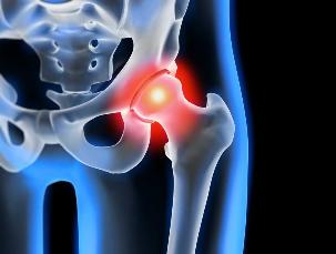
Osteoarthritis of the hip joint degenerative pathology, which is characterized by destruction of the hyaline cartilage. The disease develops gradually, associated with pain and reduced range of motion. In the absence of medical intervention at an early stage of osteoarthritis a couple of years later, there is atrophy of the thigh muscles. The injured limb was shortened, and the fusion of the joint space leads to partial or complete paralysis of the hip joint. It causes pathology gets over the trauma, curvature of the spine, systemic diseases of the muscular-skeletal system.
Osteoarthritis is typically diagnosed in patients of middle age and older people. The diagnosis put on the basis of results of instrumental investigations, x-ray, MRI, CT scan, arthroscopy. The treatment of the diseases 1 and 2, the degree of severity of the a conservative. Upon detection of ankylosis, or the failure of treatment with medications, surgery, surgery (arthrodesis, arthroplasty).
The mechanism of the development of the pathology of the
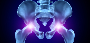
The hip joint consists of two bones — the iliac and femoral. The lower part of the Ileum (g) is its body, which is involved in the plexus to the thigh bone, which is in the upper part of the acetabulum. During the movement of the stationary glenoid fossa and the femoral head moves to the outside. The "exchange rate" with the device, the hip joint allows him to bend, stretch, rotate, contributing to the shift to the hips. The smooth gliding of the articular structures, and which provides a smooth, elastic, hyaline cartilage lining the socket and the femoral head. Its main function is the allocation of the burden of driving, which will prevent quick deterioration of bone tissue.
Under the influence of external or internal factors, and disturbed trophic of the cartilage. He does not have a vascular system — the nutrients to the tissues of the supply of synovial fluid. In the case of arthrosis it thickens and becomes viscous. Lack of nutrients causes a drying out of the surface hyaline cartilage. It is covered with cracks, which leads to a constant micro-trauma of the tissues in the flexion / extension of the hip joint. The cartilages become thinner, and lose their cushioning qualities. To "adapt" to the increase of pressure, the bones are deformed. Against the background of the deterioration of the metabolism in the tissues of the progress of the destructive-degenerative changes in the.
The cause and predisposing factors
Idiopathic or primary osteoarthritis develops without any reason. It is believed that the destruction of the cartilage due to the natural ageing of the body slows down the regenerative processes and degradation of collagen and other compounds that are necessary for the proper regeneration of the structures of the hip joint. Secondary osteoarthritis occurs on a background of the already present in the organism, a condition. The most common cause of secondary illness, including the following:
- previous injury — injury to the tendon unit, muscle tears, and their complete separation from the bone base fractures, dislocations;
- developmental disorders of the joints, congenital and dysplastic disorders;
- autoimmune diseases (rheumatoid, reactive, psoriatic arthritis, systemic lupus erythematosus;
- non-specific inflammatory diseases, such as suppurative arthritis;
- specific infections — gonorrhea, syphilis, brucellosis, anaplasmosis, trichomoniasis, tuberculosis, osteomyelitis, encephalitis;
- the disruption in the functioning of the endocrine system;
- degenerative pathology — osteochondropathy in the femoral head;
- hypermobility of the joints due to the production of a "super " capable" of collagen, which trigger their excessive mobility, the weakness of the bond.
Because it is the cause of the development of osteoarthritis may be hemarthrosis (bleeding into the cavity of the hip joint), the precipitating factors, which include, a violation of hematopoiesis. The disease, which are, excess weight, excessive exercise, a sedentary life-style. Its development will lead to a wrong organization of the training, deficiencies in the diet product with a high content of minerals, fat and water-soluble vitamins. Postoperative arthrosis occurs several years after the surgery, especially if it was accompanied by the excision of a large volume of tissue. Osteoarthritis of the hip joint cannot be transferred by inheritance. But, in the presence of certain inherent characteristics of the (a violation of metabolism, the structure of the skeleton), there is a likelihood of its development is substantially increased.
The symptoms of this
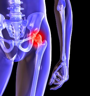
The leading symptoms of osteoarthritis of the hip joint pain when walking the thigh and the knee. A person who suffers from stiffness in the limbs, stiffness especially in the morning. For the stabilisation of the joint, and the patient begins to limp, and his gait had changed. Over a period of time, due to muscle atrophy and deformation of the joint the limb is considerably shortened. The second characteristic feature of the pathology of the — limitation of hip abduction. For example, a problem may occur when you attempt to sit up in a chair, legs apart to the sides.
Also, the "enter" ARTHRITIS can be cured at home! It's important to remember once a day to smear a it...
For arthrosis of the first stage is characterized by sporadic pain, which can occur after intense physical stress. It is located in the area of the articulation, and fade away after a long vacation.
In the second stage of the osteoarthritis of the hip joints, and the pain intensity increases. The discomfort occurs at rest, may extend to the hip and groin, worse when lifting weights or increasing physical activity. To address hip pain, a person who starts a barely noticeable limp. Marked limitation of motion of the joint, especially in abduction and internal rotation of the hip.
Arthrosis of the third stage is characterized by constant, severe pain. When driving, it is difficult, therefore, when walking, the person is forced to use a cane or a crutch. Due to the weakness of the abductor muscles of the hip joint is offset from the pelvis in the frontal plane. As a replacement for an offence to shorten the legs of the patient when the movement is tilted toward the affected limb. This leads to a shift of the centre of gravity, and the increased stress on the joint. At this stage of the arthrosis develops, marked ankylosis of the joint.
| The rate of | Radiographic signs of |
| The first | The changes are expressed not sharply. The articular ridges of moderate, irregularly constricted, there is no destruction of the surface of the femur. On the outside, or the inside edge of the acetabulum, there was a slight bony growths |
| The other | The amount of the total area of which is substantially reduced as a result of my uneven stitching. The bones of the heads of the femur is displaced upward, deformed, enlarged, and its contours become uneven. Osteophytes are formed on the surface of the inner and outer edges of the glenoid fossa of the |
| The third | There is a full or partial merger of the common room. The head of the femur is strongly expanded. More of the bone growths which are found on the surface of the acetabulum |
The diagnosis of
For the determination of a diagnosis, a physician considers clinical manifestations of the disease, anamnesis, external examination of the patient, and the instrument of the investigation. Most of the information the radiography. With its help, the estimated condition of the hip joint that determines the level of its exchange rate, the degree of damage of the cartilage tissues, and, in some cases, the cause of the development of the economy. When the cervico-diaphyseal node is increased, and the acetabulum is flattened and cut, then with high probability it can be assumed, are inherent to dysplastic changes in the joints. In Perthes disease, indicates whether the form of the femur. X-ray allows the identification and the preparation of post-traumatic osteoarthritis, despite lack of history of previous illnesses, injuries. Also, the use of other diagnostic methods:
- A CT scan helps to detect growths to the edges of the plates of bone formed osteophytes;
- An MRI was performed to assess the status of the connective-tissue structures and the extent of their participation in the pathological process.
If necessary, the inner surface of the joint is examined with the help of the arthroscopic instruments. Differential diagnosis is done to exclude osteoarthritis, lumbar-sacral or thoracic degenerative disc disease. Pain in osteoarthritis may be a masquerade, as the clinical manifestations of radicular syndrome is due to pinching or inflammation of a nerve. If you want to exclude a neurogenic pathology generally through a series of tests. Osteoarthritis of the hip joint necessary differentialsa from the top of the bursitis of the hip, ankylosing spondylitis, reactive arthritis. If you want to deactivate the auto-immune disease it is carried out biochemical studies of blood and synovial fluid.
Operation of the
With the ineffectiveness of conservative therapy to The diagnosis of complex pathology of the surgery performed. For the restoration of cartilage in joints damaged by arthritis, do surgery is impossible, but with the right approach to treatment, compliance with all health regulations, maintaining a proper way of living, medical gymnastics, and regular courses of massage, the intake of vitamins and proper diet you can stop the process of destruction, and the destruction of the cartilage of the hip joints.